New microscopy reconstructs 3D images through scattering tissue
Volumetric scattering microscopy uses information encoded in scattered fluorescence to reconstruct three-dimensional structures without mechanical scanning or wavefront measurement.
·4 min read
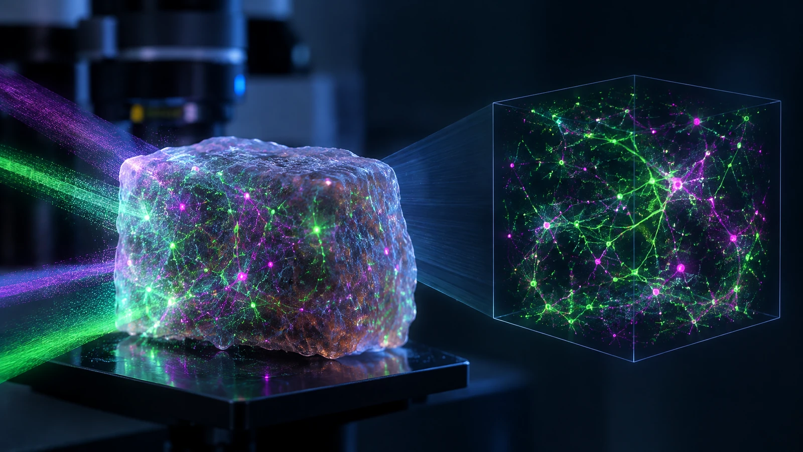
Illustration of the principle of volumetric scattering microscopy. Not the researchers' actual measurements.Illustration generated with OpenAI for Progress Radar
When tissue scrambles the light
Fluorescence microscopy can reveal cells and structures with high contrast, but biological tissue makes the task difficult. As light travels through tissue, it scatters in many directions. Some of the information about where the light came from and the direction it was travelling is therefore lost or mixed together.
The problem becomes especially important when researchers want to reconstruct a three-dimensional volume. Methods designed to compensate for scattering can require complex optics, wavefront measurements or mechanical scanning through the sample.
In a peer-reviewed Nature Communications study, researchers now present volumetric scattering microscopy, or VSM, which approaches the problem differently: it uses patterns in the scattered light as part of the information needed to reconstruct the volume.
Multiple angles in one optical setup
VSM segments the aperture in the Fourier plane so the system records fluorescence from multiple angles. The scattered light forms complex speckle patterns, but those patterns still contain information about the sample's three-dimensional structure.
A computational method analyses these data using adaptive feature-based descattering and aligns information from the different sub-apertures. The result is a reconstructed 3D representation of fluorescent structures inside the scattering material.
The researchers describe the system as scan-free. It requires neither mechanical scanning through the volume nor direct wavefront measurement, while its optical architecture builds on a comparatively simple epi-fluorescence arrangement.
Tested from model samples to intact embryos
The researchers tested VSM across several kinds of samples to determine whether the method works as scattering becomes biologically and structurally complex.
The demonstrations included artificial phantoms, engineered cellular systems, ex vivo tissue with volumetric muscle loss and intact Xenopus embryos. According to the study, the reconstructions preserved spatial resolution while improving optical sectioning and quantitative accuracy under strong scattering conditions.
That matters because a method that works in a simple transparent sample may not retain its performance when light has to pass through layered or fully embedding biological tissue. The researchers report that VSM handled both kinds of scattering environment across a large field of view.
Turning scattering into part of the signal
The central idea is that scattered light is not treated only as noise to be removed. Instead, VSM attempts to preserve the angle-dependent information that remains encoded in the scattered light and use it during reconstruction.
The method does not make tissue optically transparent, and the study does not demonstrate a finished clinical imaging system. The experiments are research demonstrations in phantoms, engineered cellular systems, tissue outside the body and Xenopus embryos.
The result is therefore best understood as a new microscopy method: a combination of comparatively simple optics and computational reconstruction that could make three-dimensional fluorescence imaging more practical in samples where light scattering would otherwise obscure the structure.
Researchers have made tiny polymer vesicles move away from a chemical source through changes in their membranes. The laboratory result reveals a simple mechanism for cell-like motion, not a working artificial cell.
Pinal translates descriptions of desired biological functions into new protein candidates. In laboratory tests, model designs showed function across four different tasks, including two active enzymes.
E(3)-Pose learns the physical symmetries of the fetal brain and estimates head position from rapid 3D MRI. The goal is to let future scanners adjust diagnostic images as the fetus moves.