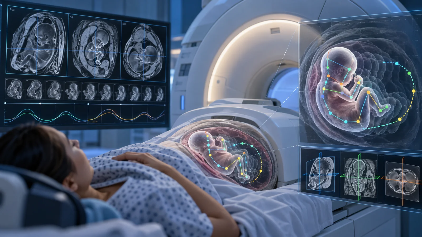
AI tracks fetal motion to make MRI scans more reliable
E(3)-Pose learns the physical symmetries of the fetal brain and estimates head position from rapid 3D MRI. The goal is to let future scanners adjust diagnostic images as the fetus moves.
·5 min read
Concept illustration of fetal MRI and AI-based motion tracking, not actual research images.Illustration: Progress Radar
A moving target inside the MRI scanner
When ultrasound raises concern about a developmental abnormality in the fetal brain, MRI can give radiologists more detailed information. But a fetus rarely stays still while the images are acquired.
Diagnostic MRI images are often collected as two-dimensional slices aligned with specific anatomical directions. If the head moves after a slice has been planned, the image can land in the wrong plane. That can make scans harder to interpret and sometimes require additional acquisition.
Researchers at MIT, Boston Children's Hospital and collaborating institutions have developed an AI method that tackles the problem before each diagnostic image is taken.
The model learns how a head can rotate
The method, called E(3)-Pose, estimates the fetal head's position and orientation in six degrees of freedom from rapid, low-resolution 3D MRI volumes.
Rather than learning every possible head position as an unrelated pattern, the model builds geometric rules into the network itself. It is designed to respond consistently to rotations and to account for anatomical symmetries that can make different orientations difficult to distinguish.
That is especially useful for clinical MRI data, which can contain noise, low resolution and artifacts. A model trained only on cleaner research images can otherwise struggle when it encounters the less predictable images seen in clinical practice.
Better generalization to clinical images
In the work, presented at ECCV 2026, the researchers compared E(3)-Pose with several existing pose-estimation strategies.
They report that the model achieved state-of-the-art accuracy on clinical MRI volumes and generalized more robustly across datasets than the methods used for comparison.
The AI is not diagnosing the fetal brain. It is trying to answer a more basic question: where is the head right now, and which direction is it facing?
An accurate answer can then be used to place the next diagnostic MRI slice in the correct orientation.
A scanner that can adjust as the fetus moves
The proposed workflow uses a rapid 3D acquisition before each diagnostic 2D image. E(3)-Pose estimates the updated head position, and that information can be used to adjust where the next slice is acquired.
The goal is a more adaptive MRI examination in which the scan follows motion instead of assuming that the fetus remains where it was at the beginning of the session.
The researchers hope this could eventually produce more reliable images, reduce manual adjustment and make examinations more comfortable by avoiding unnecessary repeat scans.
Still a research system
E(3)-Pose is not presented as a finished clinical diagnostic system. The results demonstrate pose estimation on research and representative clinical datasets and are intended to support future clinical translation.
The model is also trained primarily on third-trimester fetuses. Younger fetuses are smaller and have more room to move, making the task harder. The researchers therefore identify second-trimester imaging as an important area for further work.
What makes E(3)-Pose notable is how the AI is built: instead of relying on a large dataset to teach the model all of the relevant physics implicitly, some rules of motion and symmetry are encoded directly into the architecture.
If the method proves robust in a clinical workflow, the result could be less time spent chasing a moving fetus and more time focused on the images radiologists actually need to interpret.
Measurements in an electron-doped copper oxide directly link superconductivity to antiferromagnetic spin fluctuations. The result resolves an important part of the puzzle, but not the mechanism behind the highest transition temperatures.
Quantinuum reports 99.9995% logical memory fidelity on its Helios system. The result shows strong error suppression with a color code, while scaling to useful fault-tolerant computation remains unproven.
Researchers have developed an artificial skin with more than 10,000 touch sensors that organizes signals more like the human nervous system. In experiments, it let robots detect very fine surface structures and react automatically to potentially harmful contact.